Reference: Gray C. Case of cæsarean section in a dwarf: recovery of the mother. The British Medical Journal 1883; 2 (1189): 727.
Back to the page on Clement Frederick Gray, Frederick Clement Gray, the Grays, Robert Fyson, Ernest Last Fyson, Walter Hutchinson, the Chronicle, or The history of medical treatments, training, qualifications and regulation.
‘CASE OF CÆSAREAN SECTION IN A DWARF: RECOVERY OF THE MOTHER
BY CLEMENT GRAY, M.R.C.S., Newmarket.
IT is happily rare that such a formidable operation is required as the removal of a child from the uterus by abdominal incision; but when the pelvis is too distorted to allow the head to pass through it, and craniotomy cannot be undertaken, the Cæsarian section offers the only chance of saving the life of the mother and her child.
The following case of operation, performed under the most dis-advantageous circumstances, without antiseptic precautions, and amid the most unsanitary conditions, is probably unique of its kind, and will be read with interest by the profession [and others, then and now I would imagine, hence its inclusion on talkingdust.net].
On July 8th, 1883, at 9.30 A.M., I was requested to visit a single woman, aged 41, who was suffering from abdominal pains [interestingly this was a Sunday morning]. She was of very diminutive stature, being only four feet in height. Her head was of unusual dimensions, and the carpal extremities of the radius and ulna were much enlarged. At the age of 8 she ceased to grow; at 24 she had a severe attack of rheumatic fever, which laid her by for nine months. From that time she had led an active life, and was regarded with much sympathy by her neighbours. Although her abdomen had been getting prominent for some time, not the slightest suspicion of pregnancy was raised by her friends. Three days previous to my visit the membranes had ruptured, and there had been a drain of watery fluid from the vagina ever since. The os uteri was dilated sufficiently to admit the tip of the finger through the narrow brim of the pelvis. I came to the conclusion that she was is labour, and that the fœtus was dead. As the antero-posterior diameter of the pelvis was only three quarters of an inch, it was impossible at this visit to ascertain the nature of the presen-tation. It being evident that delivery per vias naturales would be impossible, and the patient’s strength must soon succumb if the only available remedy were not resorted to, I decided to perform Cæsarian section without further delay. With the assistance of Dr. Gray, Mr. Fyson, Mr. E. Fyson, and Mr Hutchinson of this town, I proceeded to operate at 10 P.M. [It’s interesting how late this was and over 12 hours after the initial presentation; and the degree of cooperation between practices is especially interesting – see also the friendships of Clement Gray mentioned with Walter Hutchinson in 1889 and Ernest Fyson in 1917 on the pages relating to those individuals.]
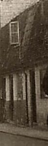
Part of an old 1940s photograph showing some even older cottages in Grosvenor Yard, likely from the time that this event took place (see below or click image for source and acknowledgements etc., ref. Image 1).
The cottage in which the patient lived is situated in a small yard, off the main street in Newmarket [Grosvenor Yard – see notes below and image on the right], and the drainage and ventilation are very defective. The bedroom in which the operation was performed was barely large enough for the four medical friends who gave me their assistance; one of them, indeed, was obliged to stand on the top of the passage-stairs. The wretched apartment was so close and the weather so sultry, that we were compelled to keep the window wide open during the operation. The patient having been placed on a common deal board, in a half-sitting posture, and the bowels and bladder having been emptied, Mr. Hutchinson proceeded to use the ether-spray. When the integu-ments of the abdomen had become insensitive to the knife, I made an incision in the line of the linea alba, commencing just below the umbilicus, and terminating within an inch of the pubes. The tissues were carefully divided down to the peritoneum; and, a director having been introduced, it was opened to the extent of the abdominal incision. The uterus now coming into view, I laid it freely open by a longitudinal incision. Both wounds (uterine and abdominal) were held together by the fingers of Mr. E. Fyson, placed within the extremities of the incision, whilst I rapidly ex-tracted the fœtus and placenta. The fœtus weighed 7 lbs., and had been dead about a week. It was grasped by both hands, and ex-tracted breech first, the head quickly following. The placenta was easily detached. The uterus contracted so closely, that the sides of the incision were soon in apposition. There was no escape of blood into the abdominal cavity, which was, therefore, only lightly sponged. Five equidistant wire sutures, including the peritoneum, brought the wound together [!], which was now covered with a com-press of lint, supported with bands of adhesive plaster and a broad bandage. The operation lasted twenty-five minutes. After the patient had been removed to her bed, a grain of opium was given by the mouth, which secured a good night’s rest.
I was amazed to find, on the following day, five children down with the measles, in the only available room for the use of the family, eight in number; the narrow stairs connecting this with the dwarf’s room above, which thus received the foul air from the lower apartment. A catheter was passed night and morning. The condi-tion of the patient for the first few days following the operation was discouraging enough; the breathing was hurried, and both pulse and temperature ran high. There was much abdominal pain and distention, and great care was necessary lest the sutures should give way. Small doses of opium and a saline mixture were prescribed. The diet consisted of milk, and from first to last no stimulant was administered. On the fifth day following the operation, the urgent symptoms began to subside; and on the eighth day the urine was passed naturally, and there was still further improvement. The bowels acted naturally on the thirteenth, when the last suture was removed. From this time, the progress towards recovery was un-interrupted; and on August 8th the wound was healed, and the patient quite well. Uterine discharge commenced immediately after the operation, and continued for three weeks. The patient had no nursing beyond that given by a neighbour, who ran in and out when she could spare time; but it is hardly necessary to say that I watched her with the closest attention.’
Note:-
Interestingly, it’s possible to identify this patient on the 1881 census, aged 38, living in Grosvenor Yard (see below also), which is just off the High Street as described. She was a boarder in the household of a groom, who had a wife and five children. In the margin she’s identified as ‘Dwarf’. On the 1891 census, the same groom with his family were living in Broughton, Salford, and the patient, by then aged 48, is identified as his sister in law. So she obviously went on to live at least another 10 years after the dramatic events described above. Reference: The National Archives, 1881 and 1891 census.
It’s also of interest that several months later, ‘Mr Clement F. Gray Medical Officer No 1 District’ applied to the Newmarket Union for some remuneration for this operation, identifying the patient by name and as ‘a dwarf pauper residing in Grosvenor Yard Newmarket’, which was granted. Reference: 18th December 1883, 611/32, Newmarket Union minutes, (Suffolk County Record Office, Bury St. Edmunds).
Image 1: From Peter Norman’s Collection; image reproduced with kind permission of Peter Norman.
Note: see comments regarding images and copyright © etc. on the Usage &c. page as well.